Get in touch
(03) 9419 6766
breastunit@breastunit.com.au

Introducing the new 3D Mammogram machine with tomosynthesis
BIV • Oct 17, 2015
A Genius 3D mammography exam is very similar to having a traditional 2D mammogram. Like a 2D mammogram, the technologist will position you, compress your breast, and take images from different angles.
During the Genius 3D mammogram, the X-ray arm sweeps in an arc over the breast, taking multiple breast images in just seconds.

There’s no additional compression required and it only takes a few seconds longer. The technologist will review the images of your breasts at the computer workstation to ensure quality images have been captured for review.
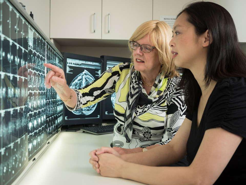
A radiologist will then examine the images and report results to either your doctor or directly to you.
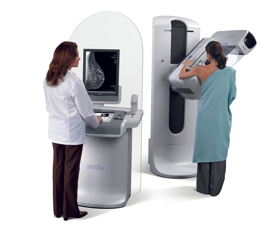
Genius 3D mammography™ allows doctors to examine your breast tissue layer by layer. So, instead of viewing all of the complexities of your breast tissue in a flat image, as with traditional 2Dmammography, fine details are more visible and significantly less likely to be hidden by the tissue above or below.
Genius 3D mammography is clinically proven to detect 41% more invasive breast cancers and reduce the need for further testing by 15-40% compared with traditional 2D mammography alone.
This means two simple things: earlier detection than ever before and less anxiety about unnecessary testing following your mammogram.

Cancer Council Victoria is reminding people how important community participation on Daffodil Day is to raise funds for cancer research, prevention programs and support services. "We want to beat cancer through more research, through educating the public on ways to prevent cancer and by helping people who have cancer get the best treatment and care." For more information on getting involved this Daffodil Day, click here .
The presentation of a pregnant or lactating woman with a palpable breast mass is a common and important scenario encountered in a breast imaging practice. The physiological changes secondary to hormonal changes result in increased breast volume and firmness, associated with nodules and increased parenchymal density. All these changes make radiological assessment of the breasts difficult. The aim of imaging in this setting is to exclude a pregnancy-associated breast cancer, which is defined as a breast cancer occurring during pregnancy or within 1 year of delivery.
Trusted for more than 30 Years

CONTACT
Level 4
21 Victoria Street
Melbourne VIC 3000
CONTACT US